真核细胞驱动蛋白马达蛋白利用 ATP 水解的能量沿细胞骨架微管网络移动货物,如染色体和囊泡 。它们在细胞内运输的方方面面都发挥着重要作用,并广泛参与各种生理过程,包括胚胎发育、轴突运输和细胞分裂。许多驱动蛋白在细胞分裂中起着重要作用,以及驱动蛋白的过度表达与癌症(如视网膜母细胞瘤)有关的事实,使它们成为开发抗有丝分裂药物非常有潜力的靶标。许多驱动蛋白仅在细胞分裂期间表达的证据也表明,它们可能优于当前的抗有丝分裂药物靶标,例如普遍表达的Tubulin。
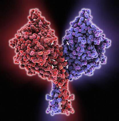
驱动蛋白的一般结构:
驱动蛋白分类和结构多样性
迄今为止,已经在从酵母(酿酒酵母 含有 6 种驱动蛋白)和真菌到人类(目前有13种驱动蛋白)等物种中鉴定了大约 150 种驱动蛋白。分类基于马达蛋白域的同源性。目前一致认为至少有九类驱动蛋白,驱动蛋白分类如表1所示,驱动蛋白命名(在表 1 中以粗体显示)基于驱动蛋白内马达蛋白结构域的位置,因此 N-蛋白质在蛋白质的氨基末端包含一个运动结构域,而 C- M-马达蛋白分别位于羧基末端和蛋白质中间。
考虑到不同类驱动蛋白的马达之间相对较高的百分比差异,很可能识别类特异性的驱动蛋白药物,这一结构证据得到了生化数据的支持,这些数据表明不同类别的果蝇驱动蛋白表现出对各种 ATP 类似物的不同的利用效率。实验表明,即使在同一的物种内,不同驱动蛋白之间也存在明显的结构差异,可用于选择性药物开发。
表 1:驱动蛋白超家族蛋白的分类及功能
| 驱动蛋白种类 | 细胞功能 | 候选HTS靶标代表 | 功能性有丝分裂抑制数据 | 参考文献 |
| N-II (BIMC) | 中心体分离 主轴动力学 (12名成员, 来自于5个门类) | AnBimC HsEg5 | 曲霉中bmc突变体抑制纺锤体分离,并引起致死表型。 在HeLa细胞中,通过显微注射HsEg5抗体导致>80%的细胞有丝分裂中止。这些结果通过过表达HsEg5马达蛋白突变体得到证实。 | 7、17、34 |
| NV (染色质) | 通过与染色体结合并帮助它们在中期板上对齐而参与细胞分裂。(7 名成员, 来自于3个门类) | 染色质驱动蛋白 | 体内反义和体外抗体抑制及免疫耗竭实验证明了染色质驱动蛋白在纺锤体组织和染色体定位中的重要作用。运动抑制导致爪蟾细胞有丝分裂阻滞和细胞死亡。人类染色体驱动蛋白的异常与视网膜母细胞瘤有关 | 18、19、20、21 |
| N-VII | 参与中期板上的染色体聚集,并可能参与后期 A 的染色体运动 (2 个成员,来自于1个门类) | HsCENP-E | 无马达 CENP-E 蛋白在人源细胞系体内过表达导致染色体无法在中期板上对齐并导致有丝分裂阻滞。此外,将 CENP-E 抗体显微注射到 HeLa 细胞中也会导致有丝分裂阻滞,持续 4 到 17 小时,并导致所有细胞进入细胞凋亡。 | 22、23、24、25 |
| N-VI (MKLP1) | 参与有丝分裂的后期 B 和胞质分裂。 (5 个成员,来自于2个门类) | HsMKLP1 | N-VI 家族的果蝇突变体表明,这种马达蛋白对于胞质分裂至关重要。 | 26、27 |
| C (C端) | 可能通过调节微管动力学参与有丝分裂和减数分裂纺锤体的形成。一些成员可能是专门的囊泡转运蛋白。 (18名成员,来自于7个门类) | HsKifC3 | 低等真核生物(酵母)中的突变分析表明,此类蛋白质中的无效突变体导致 G2/M 的致命有丝分裂阻滞。 | 35、36、37、38、39 |
| M (MCAK/KIF2 ) | 参与后期染色体运动和微管动力学。一些成员是囊泡驱动蛋白。(10名成员,来自于4个门类) | HsMCAK | 反义抑制哺乳动物的M蛋白导致后期染色体分离的中断。一个无马达蛋白突变体的过表达也导致了后期的破坏。 | 6、32、33 |
| N-I (KHC) | 细胞器/囊泡运输 (15名成员,来自于7个门类) | HsKHC | 在有丝分裂中没有作用 | 30,31 |
| N-III (Unc104) | 细胞器/囊泡运输,特别是突触囊泡和线粒体 (18名成员,来自于4个门类) | HsKIF1C | 在有丝分裂中没有作用 | 28 |
| N-Ⅳ (KRP85/95) | 细胞器/囊泡运输特别是顺行囊泡运输。 (13 名成员, 来自于4个门类) | HsKIF3C | 在有丝分裂中没有作用 | 29 |
用于 HTS 分析的驱动蛋白靶标的选择
从上表我们可以看到,驱动蛋白的细胞功能主要可分为两大类:膜囊泡和细胞器的运输和定位,以及有丝分裂纺锤体的形态发生和染色体运动 [12],九类驱动蛋白中有四类专门参与细胞分裂(表 1 的黄色/绿色阴影区域),两类(C- 和 M-)同时包含囊泡转运蛋白和有丝分裂马达蛋白(表 1 的浅黄色阴影区域),三个类别(NI、NIII 和 N-IV)仅在囊泡运输中起作用(表 1 的无阴影区域)。
我们从每个驱动蛋白类中选择了一个人类同源蛋白[6,17,21,22,26,28,29,30,39],从Aspergillus(一种人类病原体)中选择了两个真菌驱动蛋白[34], 一个人类病原体作为 HTS 检测的基础(这些显示在表 1 中)。通过用化合物库筛选每种蛋白质,可以生成一个初级化合物库,该化合物库应该允许识别选择性靶向细胞分裂特定类别的人类驱动蛋白(表 1 的深色阴影区域)的化合物,同时对囊泡转运驱动蛋白没有影响。这种化合物很有可能是治疗人类疾病如癌症的有效的抗有丝分裂剂。同样,可以将与曲霉驱动蛋白特异性反应的化合物用作潜在的抗真菌药物。
驱动蛋白为什么能作为抗有丝分裂药物筛选靶点?
在开始一项昂贵的药物开发计划之前,必须严格评估候选蛋白的适用性,以满足有效药物靶标的标准。驱动蛋白将成为抗有丝分裂药物开发的绝佳靶点的证据来自一系列实验方法:包括突变分析 [40]、抗体抑制实验 [7,32] 和驱动蛋白活性的反义抑制 [19]。这些证据总结在表 1 中,可以看出特定有丝分裂特异性驱动蛋白的功能性抑制导致细胞分裂的抑制。
在低等真核生物中,突变分析有助于阐明有丝分裂驱动蛋白的作用。单个驱动蛋白的突变通常会导致有丝分裂阻滞和致死表型。我们选择作为抗真菌靶点的 AnBimC 蛋白就是这种情况[34]。然而,在某些情况下,两个高度同源的基因执行相同的功能从而导致功能冗余[41],在这些情况下,必须同事敲除两个基因才能产生有丝分裂表型变化。
重要的是,所有数据表明,针对有丝分裂驱动蛋白的抗体会特异性影响有丝分裂过程,但对驱动蛋白囊泡转运功能没有影响 [42]。事实上,对特定驱动蛋白的抑制通常会诱导一种非常具有细胞周期特异性的表型。例如,抑制 M 类驱动蛋白 MCAK 中的运动区域对纺锤体组装没有影响,但确实抑制了染色体运动 [32]。该数据表明驱动蛋白马达的特定抑制剂将只会特异性地抑制有丝分裂过程而不影响其他关键细胞功能。
驱动蛋白靶标相对于当前抗有丝分裂靶标的优势:
微管的纺锤体蛋白Tubulin是目前抗有丝分裂药物如紫杉醇和长春碱的主要靶点 [43,44]。人们普遍认为,这些药物通过在有丝分裂期间直接抑制微管动力学发挥作用 [45,46]。由于有丝分裂细胞中的微管动力学比静止细胞大得多,因此对分裂细胞的特异性是有利的。药物处理导致有丝分裂阻滞,在此期间细胞进入凋亡途径并死亡 [47]。但由于其作用机制的性质,所有抗有丝分裂剂,包括紫杉醇和长春碱,都会在某种程度上对正常有丝分裂细胞产生不利影响,例如存在于胸腺、睾丸、小肠、结肠和胎盘中的细胞 [48] .
然而,有丝分裂驱动蛋白作为潜在的抗有丝分裂药物靶点的一个主要优点,是它们的表达通常受到严格调节,从而与有丝分裂事件一致。例如,HsMCAK 仅在增殖细胞中表达,其表达已被证明在转录水平受到严格调控 [6];HsEg5 仅在有丝分裂期间与微管相关,在体内间期与 MT 不相关 [7];染色质驱动蛋白仅在增殖细胞中表达,在那里它们是寿命较短的蛋白质,可能受细胞周期蛋白降解机制的调节 [51];研究表明,CENP-E在核破裂后立即与着丝点结合,并一直保持完全结合,直到B后期,当它重新定位到B后期纺锤体,随后通过类似周期素的途径降解 [52]。有丝分裂驱动蛋白表达的严格调控预示着这些将是高度特异性的抗有丝分裂靶点,具有最小的剂量限制副作用。
产品推荐:
在这里给大家带来一款驱动蛋白活性检测试剂盒,可用于筛选影响驱动蛋白活性的药物。
产品详情参见:驱动蛋白(Kinesin)活性检测试剂盒
| 产品名称 | 货号 |
| Kinesin ELIPA Kit | BK060 |
参考文献:
Wang, SZ & Alder, R. Chromokinesin: a DNA-binding, kinesin-like nuclear protein. J. Cell Biol. 128: 761-768 [1995]
Hughes, D. Predicting the future for R&D - science or art? Drug Disc. Today 3: 487-489 [1998]
Krantz, A. Diversification of the drug discovery process. Nature Biotech. 16: 1294 [1998]
Rao, K. Short Communication. Drug Disc. Today. 3: 349 [1998]
Ansell, J. Hot and cold areas of therapeutic R&D: A survey of the top 50 pharma companies. Mod. Drug Disc. May/June: 19-22 [1999]
Endow, S., Henikoff, S., Niedziela, L. Mediation of meiotic and early mitotic chromosome segregation in Drosophila by a protein related to kinesin. Nature 345:81-83 [1990]
Endow, S., Chandra, R., Komma, D., Yamamoto, A., Salmon, E. Mutations of the Drosophila ncd microtubule motor protein cause centrosomal and spindle pole defects in mitosis. J. Cell Sci. 107:859-867 [1994]
Meluh, P., Rose, M. KAR3, a kinesin-related gene required for yeast nuclear fusion. Cell 60:1029-1041 [1990]
O’Connell, M., Meluh, P., Rose, M., Morris, R. Suppression of the bimC4 mitotic spindle defect by deletion of klpA, a gene encoding a KAR3-related kinesin-like protein in Aspergillus nidulans. J. Cell Biol. 120:153-162 [1993]
Hoang, E., Whitehead, J., Dose, A., Burnside, B. Cloning of a novel C-terminal kinesin (KIFC3) that maps to human chromosome 16q13-q21 and thus is a candidate gene for Bardet-Biedl syndrome. Genomics 52:219-222 [1998]
Kim, I., Jun, D., Sohn, U., Kim, Y. Cloning and expression of human mitotic centromere-associated kinesin gene. Biochem. Biophys. Acta. 1359:181-186 [1997]
Maney, T., Hunter, A., Wagenbach, M., Wordeman, L. Mitotic centromere associated kinesin is important for anaphase chromosome segragation. J. Cell Biol. 142:787-801 [1998]
Waters, J., Salmon, E. Cytoskeleton: a catastrophic kinesin. Curr. Biol. 6:361-363 [1996]
Enos, A., Morris, N. Mutation of a gene that encodes a kinesin-like protein blocks nuclear division in A. nidulans. Cell 60:1019-1027 [1990]
Blangy, A., Chaussepied, P., Nigg, E. Rigor type mutation in the kinesin related protein HsEg5 changes its subcellular localization and induces microtubule bundling.
Sawin, K., LeGuellec, K., Phillippe, M., Mitchinson, T. Mitotic spindle organization by a plus end directed microtubule motor. Nature 359:540-543 [1992]
Blangy, A., Lane, H., d’Herin, P., Harper, M., Kress M., Nigg, E. Phosphorylation by p34cdc2 regulates spindle association of human Eg5, a kinesin related motor essential for bipolar spindle formation in vivo. Cell 83:1159-1169 [1995]
Walczak, C., Vernos, I., Mitchison, T., Karsenti, E. A model for the proposed roles of different microtubule-based motor proteins in establishing spindle bipolarity. Curr. Biol. 8: 903-913 [1998]
Vernos, I., Raats, J., Hirano, T., Heasman, J., Karsenti, E., Wylie, C. Xklp1, a chromosomal Xenopus kinesin-like protein essential for spindle organization and chromosome positioning. Cell 81:117-127 [1995]
Tokai, N., Fujimoto, A., Toyoshima, Y., Tsukita, S., Inoue, J., Yamamota, T. Kid, a novel kinesin-like DNA binding protein, is localized to chromosomes and the mitotic spindle. EMBO J. 15:457-567 [1996]
Yan, R.T., Wang, S.Z. Increased chromokinesin immunoreactivity in retinoblastoma cells. Gene 189:263-267 [1997]
Schaar, B., Chan, G., Maddox, P., Salmon, E., Yen, T. CENP-E function at kinetochore is essential for chromosome alignment. J. Cell Biol. 139:1373-1382 [1997]
Yen, T., Li, G., Schaar, B., Cleveland, D. CENP-E is a putitive kinetochore motor that accumulates just before mitosis. Nature 359:536-539 [1992]
Wood, K., Sakowicz, R., Goldstein, L., Cleveland, D. CENP-E is a plus end directed kinetochore motor required for metaphase chromosome alignment. Cell 91:357-366 [1997]
Lombillo, V., Nislow, C., Yen, T., Gelfand, V., McIntosh, R. Antibodies to the kinesin motor domain and CENP-E inhibit microtubule depolymerization-dependent motion of chromosomes in vitro. J. Cell Biol. 128:107-115 [1995]
Nislow, C., Lombillo, V.A., Kuriyamu, R., McIntosh, R. A plus end directed motor enzyme that moves antiparallel microtubules in vitro localizes to the interzone of mitotic spindles. Nature 359:543-547 [1992]
Adams, R.R., Tavares, A.A., Salzberg, A., Bellen, H., Glover, D. Pavoretti encodes a kinesin-like protein required to organize the central spindle and contractile ring for cytokinesis. Genes & Dev. 12:1483-1494 [1998].
Wright, B., Terasaki, M., Scholey, J. Roles of kinesin and kinesin-like proteins in sea urchin embryonic cell division: evaluation using antibody microinjection. J. Cell Biol. 123:681-689 [1993]
Puck TT. and Marcus PI. 1956. Action of X-rays on mammalian cells. J. Exp. Med. 103, 653-666.
Brown JM and Wouters BG, 1999. Apoptosis, p53, and tumor cell sensitivity to anticancer agents. Cancer Research, Rev, 59, 1391-1399.
Enos, AP & Morris, NR. Mutation of a gene that encodes a kinesin-like protein blocks nuclear division in Aspergillus nidulans. Cell 60: 1019-1027 [1990].
Yen, TJ., Li, G., Schaar, BT., Szilak, I. and Cleveland, DW. CENP-E is a putative kinetochore motor that accumulates just before mitosis. Nature 359(6395): 536-539 [1992].
Wang, SZ & Alder, R. Chromokinesin: a DNA-binding, kinesin-like nuclear protein. J.Cell Biol. 128:761-768 [1995].
Blangy, A., Lane, HA., d’Herin, P., Harper, M., Kress, M. and Nigg, EA. Phosphorylation by p34cdc2 regulates spindle association of human Eg5, a kinesin-related motor essential for bipolar spindle formation in vivo. Cell 83(7): 1159 [1995].
Navonne, F., Niclas, J., Hom-Booher, N., Sparks, L., Bernstein, H., McCaffrey, G and Vale, R. Cloning and expression of a human kinesin heavy chain gene: Interaction of the C-terminal domain with cytoplasmic microtubules in transfected CV-1 cells. J. Cell Biol. [1992].
Telford, EA., Wightman, P., Leek, J., Markham, AF., Lench, NJ. and Bonthron, DT. cDNA cloning, genomic organization, and chromosomal localization of a novel human gene that encodes a kinesin-related protein highly similar to mouse Kif3C. Biochem. Biophys. Res. Comm. 242(2): 407 [1998].
37)Hoang, E., Whitehead, J., Dose, A., Burnside, B. Cloning of a novel C-terminal kinesin (KIFC3) that maps to human chormosome 16q13-q21 and thus is a canidate gene for Bardet-Biedl syndrome. Genomics 52: 219 [1998].
Kim, IG., Jun, DY., Sohn, U. and Kim, YH. Cloning and expression of human mitotic centromere-associated kinesin gene. Biochim. Biophys. Acta 1359(3): 181 [1997].
Nislow, C., Lombillo, VA., Kuriyama, R. and McIntosh, JR. A plus-end directed motor enzyme that moves antiparallel microtubules in vitrolocalizes to the interzone of mitotic spindles. Nature 359(6395): 543 [1992].
Middleton, K. and Carbon, J. (1994). Kar3-encoded kinesin is a minus-end directed motor that functions with centromere binding proteins (CBF3) on an in vitroyeast kinetochore. Natl. Acad. Sci. USA. 91:7212-7216.
Aizawa, H., Sekine, Y., Takemura, R., Zhang, Z., Nangaku, M. and Hirokawa, N. (1992). Kinesin family in murine central nervous system. Cell Biol.119:1287-1296.
Sekine, Y. et al. (1994) A novel microtubule-based motor protein (KIF4) for organelle transports, whose expression is regulated developmentally. Cell Biol.127:187-201.
Enos, A.P. and Morris, N.R. (1990). Mutation of a gene that encodes a kinesin-like protein blocks nuclear division in A. nidulans. Cell60:1019-1027.
Nislow, C., Lombillo, V.A., Kuriyama, R. and McIntosh, J.R. (1992). A plus-end directed motor enzyme that moves antiparallel microtubules in vitrolocalizes to the interzone of mitotic spindles. Nature 359:543-547.
Vale, R.D., Reese, T.S. and Sheetz, M.P. (1985) Identification of a novel force generating protein, kinesin, involved in microtubule-based motility. Cell42: 39-50.
Pesavento, P.A., Stewart, R.J. and Goldstein, L.S.B. (1994). Characterization of the KLP68D kinesin-like protein in Drosophila: possible roles in axonal transport. Cell Biol.127: 1041-1048.
Wood, K., Sakowicz, R., Goldstein, L., Cleveland, D. CENP-E is a plus end-directed kinetochore motor required for metaphase chromosome alignment. Cell 91:357-366 [1997].
Ames, B.N., McCann, J. & Yamasaki, E. (1975) Methods for detecting carcinogens and mutagens with Salmonella/mammalian-microsome mutagenicity test. Mutation Res. 31, 347-364.
Shrivastava, R., John, G. W., Rispat, G., Chevalier, A., and Massingham, R. 1991. Can the in vivo maximum tolerated dose be predicted using in vitro techniques? A working hypothesis. ATLA 19: 393-402.
Berry, M.N. & Friend, D.S. (1969) High-yield preparation of isolated rat liver parenchymal cells. Journal of Cell Biology, 43, 506-520.
Schmetz, E.G., Hazelton, G.A., Hall, J., Watkins, P.B., Klaassen, C.D., & Guzelian, P.S. (1986). Induction of digitoxigenin monodigitoxoside UDP-glucuronyltransferase activity by glucocorticoids and other inducers of cytochrome P-450p in primary monolayer cultures of adult rat hepatocytes and in human liver. J. Biol. Chem., 261, 8270-8275.
作为一家具有高端的技术实力、先进的经营管理水平和完善的市场销售体系的生物高科技企业,总部位于武汉光谷高新技术开发区,服务面向全国。艾美捷科技是集进口试剂、实验室耗材销售、技术服务与合约开发为一体的专业化高科技公司,为用户提供专业的前沿资讯、完备的产品、整合的解决方案,及优质的物流服务。为了更好的服务客户,公司组建了一支经验丰富的研发团队-艾美捷生物技术中心,进入研发生产阶段,将更优质的产品推荐给国内生物领域的同仁们!
艾美捷科技与国内外优秀的生物试剂供应商优保持着密切的合作关系,目前已成为众多国际知名品牌的中国总代理或一级代理,主要包括:AmyJet、AAT Bioquest、Abbexa、Abnova、Agrisera、Anogen、Biosensis、Biovision、BioVendor、Caisson Labs、Cell Biolabs、Cytoskeleton、Demeditec、Duchefa、Epigentek、Equitech-Bio、EXBio、Fitzgerald、GeneCopoeia、Hycult Biotech、ImmunoReagents、ImmunoStep、Jackson、LifeSensors、LigaTrap、Lumiprobe、Mabtech、Matreya、Norgen Biotek、Origene、ProImmune、ProSpec、ScyTek、Solis BioDyne、SouthernBiotech、StressMarq、SySy、US Biological、TRC等,可以在短时间内为用户提供专业的前沿资讯、完备的产品及物流服务。
产品订购:sales@amyjet.com
邮政编码:430070
公司地址:武汉市洪山区光谷大道35号
光谷总部国际二期时代1栋13楼
提示:本公司所有产品仅供科研使用,不用于临床诊断。
版权所有:艾美捷科技有限公司 鄂ICP备10204150号-1 鄂公网安备:42018502004523号
第二类医疗器械经营备案凭证:鄂汉药监械经营备20234324号

微信扫码在线客服