艾美捷科技,Cell Biolabs中国区总代理
| 产品名称 | 货号 | 规格 | 说明 |
Comet Assay Kits, 3-Well (3孔玻片)
(最常用) | STA-350 | 15 assays(5片) | 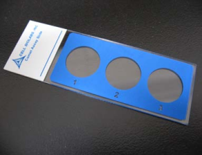
|
| STA-351 | 75 assays(25片) |
| STA-351-5 | 5*75 assays(5*25片) |
Comet Assay Kits, 96-Well(96孔玻片)
(高通量筛选) | STA-355 | 96 assays(1片) | 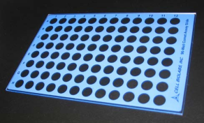
|
| STA-355-5 | 5*96 assays(5片) |
*为方便客户使用试剂盒,艾美捷科技善意将其翻译成中文操作手册,请以试剂盒中配套的英文版为准。因编者翻译水平有限,对于由本说明书中不当翻译所造成的损失,概不负责,如有错误,欢迎指正!
本说明书对应英文链接:https://www.cellbiolabs.com/sites/default/files/STA-350-comet-assay-kit.pdf
实验开始前,建议先通读说明书(请以试剂盒中配套的英文版为准):
介绍:由于环境因素和细胞内正常的新陈代谢过程,DNA损伤每天以每个细胞1000到100万个分子损伤的速度发生。虽然这只占人类基因组约60亿个碱基(30亿个碱基对)的一小部分,但关键基因的未修复损伤可能会阻碍细胞发挥其功能的能力,并显著增加癌症的可能性。彗星实验(Comet assay)也被称为单细胞凝胶电泳实验(Single cell gel electrophoresis,SCGE),是一种超灵敏的单细胞水平上检测DNA损伤的技术。在电泳场下,损伤的细胞DNA(包含碎片和链断裂)从完整的DNA中分离出来,在显微镜下形成典型的“彗星尾巴”形状。DNA损伤的程度通常是通过测量彗星尾巴来目测的,也可以使用图像分析软件来测量各种参数。
OxiSelect™ Comet Assay是一种快速、灵敏的,用于测量细胞DNA损伤的试剂盒。每个试剂盒提供足够的试剂,最多可进行15次检测。
OxiSelect™ Comet Assay实验原理:Cell Biolabs 的 OxiSelect™ Comet Assay 是一种单细胞凝胶电泳法 (SCGE),用于简单评估细胞 DNA 损伤。首先,将单个细胞与低熔点琼脂混合后,再应用到OxiSelect™ Comet特制玻片上。然后,用裂解缓冲液和碱性溶液处理这些嵌入的细胞,使DNA松弛并变性。最后,在电泳槽中对进行电泳,以分离完整的DNA和受损的片段。电泳后,样品被干燥,用DNA染料染色,并通过外显荧光显微镜可视化。在这些条件下,受损的DNA(包含裂解和链断裂)将比完整的DNA迁移更多,并产生 "彗星尾 "形状(见图1)。
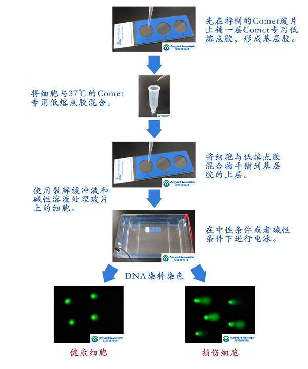
图1,彗星实验原理与流程示意图
试剂盒组分(以STA-350,3孔板为例):
| 组分 | 组分编码 | 规格 |
OxiSelect™ 3-Well Comet Slides
特制3孔玻片(表面处理) | STA-352 | 5片(每片上有3孔) |
OxiSelect™ Comet Agarose
彗星实验专用低熔点琼脂糖胶 | 235002 | 15ml(1瓶,无菌) |
Vista Green DNA Dye, 10000X
绿色荧光DNA染料,10000X | 235003 | 5ul(1支) |
EDTA Solution, 500 mM
EDTA溶液,500mM | 235004 | 50ml(1瓶) |
10X Lysis Solution
10X 裂解液 | 235005 | 20ml(1瓶) |
客户自备材料:
1.氯化钠粉末
2.NaOH颗粒
3.10 N NaOH,用于调整pH值 (10 N NaOH = 10 M NaOH =10 mol/L NaOH)
4.DMSO(可选)
5.70%乙醇
6.TE缓冲液(10mM Tris,pH 7.5,1mM EDTA)。
7.PBS(不含Mg2+和Ca2+)。
8.EDTA(二钠盐)
9.DI H2O(去离子水)
10.37ºC和沸水浴
11.水平电泳槽
12.可调节的单通道微量移液器,带一次性吸头。
13.带FITC过滤器的外显荧光显微镜
保存条件:收到试剂盒后,将绿色荧光DNA染料,10000X存放在-20ºC。 所有其他试剂盒组件在室温下储存。
《实验步骤》(以STA-350,3孔板为例)
为方便客户使用试剂盒,艾美捷科技善意将其翻译成中文操作手册,请以试剂盒中配套的英文版为准。因编者翻译水平有限,对于由本说明书中不当翻译所造成的损失,概不负责,如有错误,欢迎指正!本说明书对应英文链接:https://www.cellbiolabs.com/sites/default/files/STA-350-comet-assay-kit.pdf
一、试剂准备:
1.OxiSelect™ CometAgarose:将Comet Agarose瓶放在90-95ºC水浴中加热20分钟,或者直到琼脂胶液化。将瓶子转移到37℃的水浴中20分钟,并保持直到使用。
2.1X Vista Green DNA Dye工作液:使用TE缓冲液(10 mM Tris, pH 7.5, 1 mM EDTA)稀释原10000X的Vista Green DNA Dye母液,按照1:10000的比例,稀释成1X的Vista Green DNA Dye工作液。1X工作液可在4℃避光条件下保存3周。
3.Lysis Buffer: 准备100ml的1X 裂解缓冲液
| NaCl | 14.6 g |
| EDTA Solution (试剂盒提供) | 20ml |
| 10X Lysis Solution (试剂盒提供) | 10ml |
| DMSO | 10ml(样品中含血红素,则加入) |
| DI H2O(去离子水) | 调整体积到90ml |
| 充分混合以溶解NaCl。缓慢加入10 M的NaOH来调节Lysis Buffer的pH值到10.0,最后再使用去离子水调整体积,定容到100ml。冷藏裂解液到4℃,直到使用。 |
| * Note: 缓冲液在室温下会看起来浑浊,但在4ºC时会变清。pH也会保持在约10.0。 |
* 血红素在4℃成胶状,用DMSO溶解的血红素。DMSO的添加是任选的,仅对含有血红素的样品(例如血细胞或组织样品)是必需的。加入DMSO对实验结果没有影响。
4.Alkaline Solution:准备100ml的碱性溶液
| NaOH | 1.2g |
| EDTA Solution (试剂盒提供) | 0.2ml |
| DI H2O(去离子水) | 调整体积到100ml |
| 充分混合以溶解NaOH。冷藏碱性溶液到4℃,直到使用。 |
5.Electrophoresis Running Solution: 准备1L电泳缓冲液,需求选择电泳模式(二选一):
| 电泳模式 | 检测类型 | 电泳时间 | 说明 |
| TBE电泳(中性电泳) | 检测单链和双链DNA的断裂 | 推荐
10-15 mins | 分析细胞凋亡的首选方法,可使用尾巴长度而非尾矩进行数据分析。 |
| 碱性电泳 | 检测单链和双链DNA的断裂,AP位点损伤在内的所有DNA 损伤 | 推荐
15-30mins | 灵敏度最高! |
* 实验过程中的各类缓冲液都需要4℃预冷。
5.1 TBE Electrophoresis Solution (中性)
| Tris Base | 10.8g |
| Boric Acid | 5.5g |
| EDTA(二钠盐) | 0.93g |
| DI H2O(去离子水) | 调整体积到1L |
| 充分混合以溶解固体颗粒。冷藏TBE电泳缓冲液到4℃,直到使用。 |
或者5.2 Alkaline Electrophoresis Solution (300 mM NaOH, pH >13, 1 mM EDTA)(碱性)
| NaOH | 12g |
| EDTA Solution (试剂盒提供) | 2ml |
| DI H2O(去离子水) | 调整体积到1L |
| 充分混合以溶解NaOH。冷藏碱性电泳缓冲液到4℃,直到使用。 |
特殊说明:为避免紫外线对细胞样品造成额外损害,请在弱光条件下进行实验。
二、准备样品和玻片:
1.准备裂解缓冲液、碱性溶液和电泳缓冲液(见试剂准备),然后再进行实验。将所有溶液彻底冷却至4ºC。
2.将OxiSelect™ Comet Agarose放在90-95ºC水浴中加热20分钟,或者直到琼脂胶液化。将瓶子转移到37℃的水浴中20分钟进行冷却,并保持直到使用。
3.在OxiSelect™ Comet玻片上,每孔加入75 ul 37℃的Comet Agarose,形成基层胶。用移液器将胶溶液涂抹在孔上,确保完全覆盖和平铺。将载玻片转移到4ºC下水平放置15分钟,使其固化。
4.按照如下方式准备细胞样品,与对照细胞:
悬浮细胞:以700×g离心2mins,弃上清,用冰冷的PBS(无Mg2+和Ca2+)洗涤细胞一次,离心,弃上清。最后,用预冷4℃的PBS(无Mg2 +和Ca 2 +)重悬细胞到1×10⁵个细胞/ml 。
贴壁细胞:轻轻地从细胞培养瓶/培养皿中取出细胞,用橡胶细胞刮处理。将细胞悬浮液转移到锥形管(EP)中,并在700×g离心2mins,弃上清。用冰冷的PBS(不含Mg2+和Ca2+)洗涤细胞一次,离心,并丢弃上清液。最后,用预冷4℃的PBS(无Mg2 +和Ca 2 +)重悬细胞到1×10⁵个细胞/ml 。
组织样品:使用解剖剪刀,切碎一小块组织到1-2ml冰冷的含20mM EDTA的PBS缓冲液(无Mg2 +和Ca 2 +)。组织/细胞悬浮液放置5分钟,然后将上清液转移到离心管中,避免转移碎片。700×g离心2mins,丢弃上清液。最后,用预冷4℃的PBS(无Mg2 +和Ca 2 +)重悬细胞到1×10⁵个细胞/ml 。
* 推荐阳性对照:使用20 uM Etoposide(依托泊苷)处理4小时。
5.将细胞悬液与Comet Agarose(步骤2)以1:10的比例(v/v),用移液枪混合均匀,并立即转移75ul 到每个孔中的原基层胶上方(步骤3)。确保完全覆盖平铺孔板,非常轻柔和小心地用移液管尖传播的悬浮液,而不干扰基层胶。
Note: 对于多个样品的处理,将细胞与Comet Agarose混合液保持在37℃,避免凝胶化。准备好样品后,重新混匀,依次取出75ul加入到玻片孔中。
想要获取完整彗星分析中文说明书文档,
请扫码下方二维码,联系专属客服获取哦~

发表文献
1.Siemionow, M. et al. (2020). Transplantation of Dystrophin Expressing Chimeric (DEC) Human Cells of Myoblast/MSC Origin Improves Function in Duchenne Muscular Dystrophy Model. Stem Cells Dev. doi: 10.1089/scd.2020.0161.
2.Lammert, C.R. et al. (2020). AIM2 inflammasome surveillance of DNA damage shapes neurodevelopment. Nature. doi: 10.1038/s41586-020-2174-3.
3.Shibayama, Y. et al. (2020). Aberrant (pro)renin receptor expression induces genomic instability in pancreatic ductal adenocarcinoma through upregulation of SMARCA5/SNF2H. Commun Biol. 3(1):724. doi: 10.1038/s42003-020-01434-x.
4.Han, J. et al. (2020). Elevated CXorf67 Expression in PFA Ependymomas Suppresses DNA Repair and Sensitizes to PARP Inhibitors. Cancer Cell. doi: 10.1016/j.ccell.2020.10.009.
5.Hays, E. et al. (2020). The SWI/SNF ATPase BRG1 stimulates DNA end resection and homologous recombination by reducing nucleosome density at DNA double strand breaks and by promoting the recruitment of the CtIP nuclease. Cell Cycle. doi: 10.1080/15384101.2020.1831256.
6.Ibnu Rasid, E.N. et al. (2020). Effect of Dioscorea hispida var. Daemona (Roxb) Prain & Burkill on Oxidative Stress and DNA Damage in the Liver of Pregnant Rats. Int J Biomed Sci. 16(3).
7.Le, B.V. et al. (2020). TGFβR-SMAD3 Signaling Induces Resistance to PARP Inhibitors in the Bone Marrow Microenvironment. Cell Rep. 33(1):108221. doi: 10.1016/j.celrep.2020.108221.
8.Klotz-Noack, K. et al. (2020). SFPQ Depletion Is Synthetically Lethal with BRAFV600E in Colorectal Cancer Cells. Cell Rep. 32(12):108184. doi: 10.1016/j.celrep.2020.108184.
9.Ito, S.S. et al. (2020). Inhibition of the ATR kinase enhances 5-FU sensitivity independently of non-homologous end-joining and homologous recombination repair pathways. J Biol Chem. doi: 10.1074/jbc.RA120.013726.
10.Klak, M. et al. (2020). Irradiation with 365 nm and 405 nm wavelength shows differences in DNA damage of swine pancreatic islets. PLoS One. 15(6):e0235052. doi: 10.1371/journal.pone.0235052.
11.Khalil, A.M. et al. (2020).  Association between Mobile Phone Using and DNA Damage of Epithelial Cells of the Oral Mucosa. J Biotechnol Biomed. 3(2020): 50-66. doi: 10.26502/jbb.2642-91280027.
12.Wang, Y. et al. (2020). Targeting therapeutic vulnerabilities with PARP inhibition and radiation in IDH-mutant gliomas and cholangiocarcinomas. Sci Adv. doi: 10.1126/sciadv.aaz3221.
13.Fang, Y. et al. (2020). Epigenetic dysregulation of Mdr1b in the blood-testis barrier contributes to dyszoospermia in mice exposed to cadmium. Ecotoxicol Environ Saf. 190:110142. doi: 10.1016/j.ecoenv.2019.110142.
14.Cupello, S. et al. (2019). Distinct roles of XRCC1 in genome integrity in Xenopus egg extracts. Biochem J. 476(24):3791-3804. doi: 10.1042/BCJ20190798.
15.Naci, D. et al. (2019). Cell adhesion to collagen promotes leukemia resistance to doxorubicin by reducing DNA damage through the inhibition of Rac1 activation. Sci Rep. 9(1):19455. doi: 10.1038/s41598-019-55934-w.
16.Lu, S. et al. (2019). Additive effects of a small molecular PCNA inhibitor PCNA-I1S and DNA damaging agents on growth inhibition and DNA damage in prostate and lung cancer cells. PLoS One. 14(10):e0223894. doi: 10.1371/journal.pone.0223894.
17.Dedobbeleer, M. et al. (2019). MKP1 phosphatase is recruited by CXCL12 in Glioblastoma cells and plays a role in DNA Strand Breaks Repair. Carcinogenesis. doi: 10.1093/carcin/bgz151.
18.Zhou, J. et al (2019). Aberrantly Expressed Timeless Regulates Cell Proliferation and Cisplatin Efficacy in Cervical Cancer. Hum Gene Ther. doi: 10.1089/hum.2019.080.
19.Sarkar, R. et al. (2019). Rotavirus activates a noncanonical ATM-Chk2 branch of DNA damage response during infection to positively regulate viroplasm dynamics. Cell Microbiol. doi: 10.1111/cmi.13149.
20.Maner, J. et al. (2019). Hexachlorobenzene exerts genotoxic effects in a humpback whale cell line under stable exposure conditions. RSC Adv. 9:39447-39457. doi: 10.1039/C9RA05352B.
21.Han, Y. et al. (2019). Exposure to waterborne nTiO2 reduces fertilization success and increases polyspermy in a bivalve mollusc: A threat to population recruitment. Environ Sci Technol. doi: 10.1021/acs.est.9b03675.
22.Shansky, Y.D. et al. (2019). Human Platelet Lysate Sustains the Osteogenic/Adipogenic Differentiation Potential of Adipose-Derived Mesenchymal Stromal Cells and Maintains Their DNA Integrity in vitro. Cells Tissues Organs. doi: 10.1159/000502813.
23.Gothai, S. et al. (2019). In Vitro and In Vivo-Scientific Evaluation on Cytotoxicity and Genotoxicity of Traditional Medicinal Plant Couroupita Guianensis Aubl. Flower. PhOL. 2:24-38. 
24.Al Khateeb, W. et al. (2019). Growth, Yield and Genetic Integrity of Spinach and Chrysanthemum as Affected by Soil Supplementation with Dam Sediments Collected From King Talal and Al-Mujib Dams/ Jordan. World Appl. Sci. J. 37(1):58-69. doi: 10.5829/idosi.wasj.2019.58.69.
25.Su, J. et al. (2019). Genomic Integrity Safeguards Self-Renewal in Embryonic Stem Cells. Cell Rep. 28(6):1400-1409.e4. doi: 10.1016/j.celrep.2019.07.011.
26.Xia, Y. et al. (2019). Rescue of DNA damage after constricted migration reveals a mechano-regulated threshold for cell cycle. J Cell Biol. pii: jcb.201811100. doi: 10.1083/jcb.201811100.
27.Chesnokova, V. et al. (2019). Growth hormone induces colon DNA damage independent of IGF-1. Endocrinology. pii: en.2019-00132. doi: 10.1210/en.2019-00132.
28.Garzón, J. et al. (2019). Human RIF1-Protein Phosphatase 1 Prevents Degradation and Breakage of Nascent DNA on Replication Stalling. Cell Rep. 27(9):2558-2566.e4. doi: 10.1016/j.celrep.2019.05.002.
29.Nakashima, A. et al. (2019). Autophagy is a new protective mechanism against the cytotoxicity of platinum nanoparticles in human trophoblasts. Sci Rep. 9(1):5478. doi: 10.1038/s41598-019-41927-2.
30.Pillay, N. et al. (2019). DNA Replication Vulnerabilities Render Ovarian Cancer Cells Sensitive to Poly(ADP-Ribose) Glycohydrolase Inhibitors. Cancer Cell. 35(3):519-533.e8. doi: 10.1016/j.ccell.2019.02.004.
作为一家具有高端的技术实力、先进的经营管理水平和完善的市场销售体系的生物高科技企业,总部位于武汉光谷高新技术开发区,服务面向全国。艾美捷科技是集进口试剂、实验室耗材销售、技术服务与合约开发为一体的专业化高科技公司,为用户提供专业的前沿资讯、完备的产品、整合的解决方案,及优质的物流服务。为了更好的服务客户,公司组建了一支经验丰富的研发团队-艾美捷生物技术中心,进入研发生产阶段,将更优质的产品推荐给国内生物领域的同仁们!
艾美捷科技与国内外优秀的生物试剂供应商优保持着密切的合作关系,目前已成为众多国际知名品牌的中国总代理或一级代理,主要包括:AmyJet、AAT Bioquest、Abbexa、Abnova、Agrisera、Anogen、Biosensis、Biovision、Caisson Labs、Cayman Chemical、Cell Biolabs、Cytoskeleton、Demeditec、Duchefa、Epigentek、Equitech-Bio、EXBio、Fitzgerald、GeneCopoeia、Hycult Biotech、ImmunoReagents、Jackson、LifeSensors、LigaTrap、Mabtech、Matreya、Norgen Biotek、Origene、ProImmune、ProSpec、ScyTek、Solis BioDyne、SouthernBiotech、StressMarq、SySy、US Biological、TRC 等,可以在短时间内为用户提供专业的前沿资讯、完备的产品及物流服务。