造影剂(又称对比剂,contrast media)是为增强影响观察效果而注入(或服用)到人体组织或器官的化学制品。这些制品的密度高于或低于周围组织,形成的对比用某些器械显示图像。如X线观察常用的碘制剂、硫酸钡等。
造影剂是介入放射学操作中最常使用的药物之一,主要用于血管、体腔的显示。造影剂种类多样,目前用于介入放射学的造影剂多为含碘制剂。自1924年国用50%的碘化钠成功地作了第一例股动脉造影以来,与介入放射学的发展一样,造影剂产品不断的更新换。
今天给大家带来的是艾美捷代理品牌Nanoprobes厂家生产的两款无与伦比的金颗粒X射线造影剂:AuroVist 15 nm&AuroVist 1.9 nm。
AuroVist 15 nm&AuroVist 1.9 nm
AuroVist特点:
高对比度&分辨率:相较于任何血池造影剂有最高的对比度;
血液中半衰期长:相较于任何造影剂,有最长的血液保留时间- 24 小时!
低毒性:AuroVist 15 nm的LD50 >5.0 g Au/kg,AuroVist 1.9nm的LD50 >1.4 g Au/kg,生物相容性高;
低渗透压:即使在高浓度下依旧保持低渗透压
低粘稠度:类似于水;易于注射,甚至可以注入小血管
非常适合microCT 成像
可浓缩至600 mg Au/mL 以获得超强对比度
将200 mg Au/mL 注射到小鼠体内会在血管中产生 1800 HU对比值
AuroVist相较于传统碘造影剂的优势:
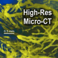
AuroVist具有非常低的粘度和渗透性,因此可以在小血管中注射和使用,而没有血管损伤的风险。在活体动物身上成功地用显微CT成像了20um血管,并首次获得了血管模型;
半衰期长:肾脏精细结构可在注射后一小时或更长时间成像;在肾脏中的浓度可提供高达1500 HU或更高的对比值
AuroVist显示出低急性毒性,更适合于药代动力学研究;
AuroVist 1.9nm分子小,可透过肾脏。
AuroVist使用剂量指南:每 40mg小瓶可供多少只小鼠使用?
一般对比度:每40 mg 小瓶 10 只小鼠一般使用效果很好。
高对比度:每40 mg 小瓶 2 只小鼠产生非常高的对比度。星云尼等人。在 2013 年的比较研究中使用此剂量,发现衰减为 2.33/mm,比同等剂量的其他 X 射线造影剂要好得多。
超分辨率:每只鼠标使用整个40 mg 小瓶。这是Hainfeld 等人 2013 年论文中使用的剂量。在我们的实验室中,即使是非常高的剂量,我们也没有发现毒性问题——到目前为止,我们只受到小鼠可以服用的总体积的限制。
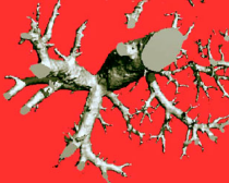
AuroVist 15 nm成像结果展示:
|
|
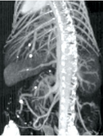
| 注射AuroVist 15 nm X射线造影剂后小鼠体内显微CT。 | 静脉注射AuroVist-15nm后,活体小鼠腿部和骨盆区域周围的20um血管显微CT。 |
| |
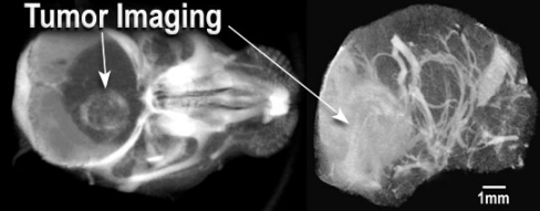
| 静脉注射AuroVist-15nm后活小鼠大脑的显微CT: 左:由于AuroVist-15nm的穿透,肿瘤突出(白色)通过受损的血脑肿瘤屏障。 右:通过计算去除头骨,显示大脑血管系统。 | |
AuroVist 1.9 nm成像结果展示:
| |
| 静脉注射AuroVist 1.9nm后小鼠肾脏的显微CT。 | 活鼠注射AuroVist x射线造影剂后5分钟成像。 |
| 活体小鼠静脉注射AuroVist 1.9nm后的肾脏成像: 我们可以发现:小的纳米金颗粒从血液中过滤出来,通过肾小球、肾小管进入输尿管。 | |
| |
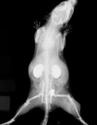
| 小鼠下腔静脉显微CT。 | 小鼠后腿体内x线片及注射AuroVistTM 1.9nm。右腿可见皮下肿瘤,血管金增多。 |
AuroVist 造影结果视频展示:
| 小鼠显微CT,静脉注射AuroVist-15nm后4小时:注意到血液和心脏中的对比度非常高,肝脏中的积聚很少。AuroVist 是血管可视化的一个革命性工具——它可以提供3倍于碘的对比度,而且不会造成血管损伤! |
| AuroVist是显微CT的完美造影剂,经多项独立对比研究证实是市场上最好的造影剂。无毒AuroVist 15 nm是一种金x射线造影剂,它有比任何其他血液池造影剂都高的对比度,并且是所有血液造影剂中最长的半衰期最长的(24小时)。由于AuroVist无毒,您可以在造影后对动物进行再处理和再扫描,以研究癌症或血管疾病的进展情况。目前已经被首次证实可用于见证主动脉瘤的发展!由于具有类似于水的稠度,AuroVist很容易注射入血管,甚至可以进入最小的血管,从而完全可视化血管系统。它会优先积聚在血管生成组织(包括肿瘤)中,用于突出显示癌症、炎症和其他血管疾病的影像。AuroVist累积已被确立为癌症生长的一种测量工具。 |
| AuroVist是显微CT的完美造影剂,经多项独立对比研究证实是市场上最好的造影剂。无毒AuroVist 15 nm是一种金x射线造影剂,它有比任何其他血液池造影剂都高的对比度,并且是所有血液造影剂中最长的半衰期最长的(24小时)。由于AuroVist无毒,您可以在造影后对动物进行再处理和再扫描,以研究癌症或血管疾病的进展情况。目前已经被首次证实可用于见证主动脉瘤的发展!由于具有类似于水的稠度,AuroVist很容易注射入血管,甚至可以进入最小的血管,从而完全可视化血管系统。它会优先积聚在血管生成组织(包括肿瘤)中,用于突出显示癌症、炎症和其他血管疾病的影像。AuroVist累积已被确立为癌症生长的一种测量工具。 |
| AuroVist 1.9nm金纳米粒子造影剂给药后小鼠肾脏切片的显微CT。AuroVist-1.9nm,因为它小于3nm,所以主要通过肾脏过滤和清除,从而提供肾脏精细结构的高对比度、高分辨率成像。 |
相关产品推荐:
| 货号 | 名称 | 规格 |
| 1115-40MGAu | AuroVist 15 nm | 40mg |
| 1115A-5X40MGAu | AuroVist 15 nm | 5x40mg |
| 1102-40MGAu | AuroVist 1.9 nm | 40mg |
| 1102A-5X40MGAu | AuroVist 1.9 nm | 5x40mg |
AuroVist引文赏析(按研究目的分类):
AuroVist与其他x射线造影剂比较研究:
Laura Nebuloni, Gisela A. Kuhn, Ralph Müller. A Comparative Analysis of Water-Soluble and Blood-Pool Contrast Agents for in vivo Vascular Imaging with Micro-CT.Academic Radiology 2013; 20:1247–1255.
Connor A. Wathen, Nathan Foje, Tony van Avermaete, Bernadette Miramontes, Sarah E. Chapaman, Todd A. Sasser, Raghuraman Kannan, Steven Gerstler, and W. Matthew Leevy. In vivo X-Ray Computed Tomographic Imaging of Soft Tissue with Native, Intravenous, or Oral Contrast.Sensors (Basel). 2013 June; 13(6): 6957–6980.
Hrvoje Lusic and Mark W. Grinstaff. X-ray-Computed Tomography Contrast Agents. Rev. 2013, 113, 1641-1666.
Assessing the toxicity of gold nanoparticles in vitro and in vivoY Pan-Bartneck - 2011 - darwin.bth.rwth-aachen.de
Hainfeld, J. F.; Slatkin, D. N.; Focella, T. M, and Smilowitz, H. M.: Gold nanoparticles: a new X-ray contrast agent. J. Radiol., 79, 248-253 (2006).
血管生成/动脉瘤/肿瘤成像:
Liu J, Fan W, Liu M, Lin X, Wang Y, Wang F, Chen X, Cao F, Liang J. Spatial Vascular Volume Fraction Imaging for Quantitative Assessment of Angiogenesis.Mol Imaging Biol. 2013 Oct 25.
Bols, J., Degroote, J., Trachet, B., Verhegghe, B., Segers, P., Vierendeels, J. A computational method to assess the in vivo stresses and unloaded configuration of patient-specific blood vessels.Journal of Computational & Applied Mathematics. July 2013, Vol. 246, p10-17.
Clark, D. P.; Ghaghada K.; Moding E. J.; Kirsch D. G., and Badea, C. T.: In vivo characterization of tumor vasculature using iodine and gold nanoparticles and dual energy micro-CT. Med. Biol., 58, 1683-1704 (2013).
SJ Tu, PY Yang, JH Hong, CJ Lo. Quantitative dosimetric assessment for effect of gold nanoparticles as contrast media on radiotherapy planning.Radiation Physics and Chemistry, 2013.
James F Hainfeld, Henry M Smilowitz, Michael J O'Connor, Farrokh Avraham Dilmanian & Daniel N Slatkin. Gold nanoparticle imaging and radiotherapy of brain tumors in mice.NanomedicineOctober 2013, Vol. 8, 10, Pages 1601-1609.
Ricketts, K.P.M.; (2012) Nanoparticles for tumour diagnostics.Doctoral thesis, UCL (University College London).
Trachet, B.; Renard, M.; de Santis, G.; Staelens, S.; de Backer, J.; Antiga, L.; Loeys, B., and Segers, P.: An integrated framework to quantitatively link mouse-specific hemodynamics to aneurysm formation in angiothensin II-infused ApoE-/- Mice. Biomed. Eng., 39, 2430–2444 (2011).
金增强放射治疗(癌症治疗):
James F Hainfeld, Henry M Smilowitz, Michael J O'Connor, Farrokh Avraham Dilmanian & Daniel N Slatkin. Gold nanoparticle imaging and radiotherapy of brain tumors in mice.NanomedicineOctober 2013, Vol. 8, 10, Pages 1601-1609.
James F Hainfeld, F Avraham Dilmanian, Zhong Zhong, Daniel N Slatkin, John A Kalef-Ezra and Henry M Smilowitz. Gold nanoparticles enhance the radiation therapy of a murine squamous cell carcinoma.2010 Med. Biol. 553045.
Hainfeld, J. F., Slatkin, D. N., and Smilowitz, H. M.: The use of gold nanoparticles to enhance radiotherapy in mice. Med. Biol., 49, N309-N315 (2004).
体内血管造影:
Liu J, Fan W, Liu M, Lin X, Wang Y, Wang F, Chen X, Cao F, Liang J. Spatial Vascular Volume Fraction Imaging for Quantitative Assessment of Angiogenesis.Mol Imaging Biol. 2013 Oct 25.
Bols, J., Degroote, J., Trachet, B., Verhegghe, B., Segers, P., Vierendeels, J. A computational method to assess the in vivo stresses and unloaded configuration of patient-specific blood vessels.Journal of Computational & Applied Mathematics. July 2013, Vol. 246, p10-17.
Trachet, B.; Renard, M.; de Santis, G.; Staelens, S.; de Backer, J.; Antiga, L.; Loeys, B., and Segers, P.: An integrated framework to quantitatively link mouse-specific hemodynamics to aneurysm formation in angiothensin II-infused ApoE-/- Mice. Biomed. Eng., 39, 2430–2444 (2011).
Hainfeld, J. F.; Slatkin, D. N.; Focella, T. M., and Smilowitz, H. M.: In Vivo Vascular Casting. Microanal., 11, (Suppl. 2: Proceedings); p. 1216CD (2005).
Micro-CT成像:
Liu J, Fan W, Liu M, Lin X, Wang Y, Wang F, Chen X, Cao F, Liang J. Spatial Vascular Volume Fraction Imaging for Quantitative Assessment of Angiogenesis.Mol Imaging Biol. 2013 Oct 25.
Laura Nebuloni, Gisela A. Kuhn, Ralph Müller. A Comparative Analysis of Water-Soluble and Blood-Pool Contrast Agents for in vivo Vascular Imaging with Micro-CT.Academic Radiology 2013; 20:1247–1255.
Bols, J., Degroote, J., Trachet, B., Verhegghe, B., Segers, P., Vierendeels, J. A computational method to assess the in vivo stresses and unloaded configuration of patient-specific blood vessels.Journal of Computational & Applied Mathematics. July 2013, Vol. 246, p10-17.
Connor A. Wathen, Nathan Foje, Tony van Avermaete, Bernadette Miramontes, Sarah E. Chapaman, Todd A. Sasser, Raghuraman Kannan, Steven Gerstler, and W. Matthew Leevy. In vivo X-Ray Computed Tomographic Imaging of Soft Tissue with Native, Intravenous, or Oral Contrast.Sensors (Basel). 2013 June; 13(6): 6957–6980.
Hrvoje Lusic and Mark W. Grinstaff. X-ray-Computed Tomography Contrast Agents. Rev. 2013, 113, 1641-1666.
Trachet, B.; Renard, M.; de Santis, G.; Staelens, S.; de Backer, J.; Antiga, L.; Loeys, B., and Segers, P.: An integrated framework to quantitatively link mouse-specific hemodynamics to aneurysm formation in angiothensin II-infused ApoE-/- Mice. Biomed. Eng., 39, 2430–2444 (2011).
Assessing the toxicity of gold nanoparticles in vitro and in vivoY Pan-Bartneck - 2011 - darwin.bth.rwth-aachen.de
Hainfeld, J. F.; Slatkin, D. N.; Focella, T. M, and Smilowitz, H. M.: Gold nanoparticles: a new X-ray contrast agent. J. Radiol., 79, 248-253 (2006).
以上产品仅用于科研,不能用于临床诊断~
看到这儿,您心动了吗?马上联系小艾吧!
或者扫描下方二维码,即可联系您的专属客服哦~

作为一家具有高端的技术实力、先进的经营管理水平和完善的市场销售体系的生物高科技企业,总部位于武汉光谷高新技术开发区,服务面向全国。艾美捷科技是集进口试剂、实验室耗材销售、技术服务与合约开发为一体的专业化高科技公司,为用户提供专业的前沿资讯、完备的产品、整合的解决方案,及优质的物流服务。为了更好的服务客户,公司组建了一支经验丰富的研发团队-艾美捷生物技术中心,进入研发生产阶段,将更优质的产品推荐给国内生物领域的同仁们!
艾美捷科技与国内外优秀的生物试剂供应商优保持着密切的合作关系,目前已成为众多国际知名品牌的中国总代理或一级代理,主要包括:AAT Bioquest、Abbexa、Abnova、ATSbio、Agrisera、Atlas Antibodies、BellBrook、Bio-Helix、Biomatik、Biosensis、BioVendor、CalBioreagents、Cayman Chem、Cell Biolabs、Columbia Biosciences、Crystal Chem、Cytoskeleton、DIAsource、Duchefa、Ebba Biotech、Echelon Biosciences、ECM Biosciences、Enzo Life Sciences、Epigentek、Equitech-Bio、FabGennix、Fitzgerald、Fluorochrome、GeneCopoeia、G-Biosciences、GroPep、Hycult Biotech、Ichorbio、Icosagen、Immundiagnostik、Immunochemistry、ImmunoReagents、Jackson、Kingfisher、KRISHGEN、LC Labs、LifeSensors、Lumiprobe、Mabtech、Matreya、Medkoo Biosciences、MyBioSource、Nanoprobe、Norgen Biotek、PeproTech、Polypure、PolySciTech、ProSci、ProSpec、ReliaTech、Rockland、SouthernBiotech、StressMarq、SySy、TRC、US Biological 等,可以在短时间内为用户提供专业的前沿资讯、完备的产品及物流服务。
产品订购:sales@amyjet.com
邮政编码:430070
公司地址:武汉市洪山区光谷大道35号
光谷总部国际二期时代1栋13楼
提示:本公司所有产品仅供科研使用,不用于临床诊断。
版权所有:艾美捷科技有限公司 鄂ICP备10204150号-1 鄂公网安备:42018502004523号
第二类医疗器械经营备案凭证:鄂汉药监械经营备20234324号

微信扫码在线客服